GENERAL INFORMATION:
History:
Nuclear Medicine history goes back to early years of 1800s when English Chemist John Dalton suggested atomic theory, 1895 when German Wilheim Konrad Roentgen found X rays and 1928 when Ernest Lawrence invented cyclotron in USA. The most important step in the development of Nuclear Medicine is the invention of artificial radioactivity by Marie Curie in 1934. However, many historians refer to 1940s when radioactive iodine was started to be used in toxic goiter as the actual date of beginning of Nuclear Medicine. Technetium, a radioactive agent that is still used most frequently in Nuclear Medicine, was artificially produced in 1937 and begun to be produced and used commercially after 1965. Different agents were found in the following years and Nuclear Medicine started to progress rapidly. The beginning of Nuclear Medicine in Turkey goes back to the end of 1960s and the first days of 1970s along with Europe and USA.
Baskent University, Department of Nuclear Medicine was established in 1995.
The department has 2 cameras: SPECT/CT and PET/CT. Other devices existing in our department are thyroid uptake device, Carbon-14 urea breath test measuring device, gamma probe and treadmill device. High dose radionuclide treatment center is available in Yapracık – Ankara.
Introduction:
Nuclear Medicine imaging uses small amounts of radioactive materials called radiotracers that are typically injected into the bloodstream, inhaled or swallowed. The radiotracer travels through the area being examined and gives off energy in the form of gamma rays which are detected by a special camera and a computer to create images. Nuclear Medicine imaging provides unique information that often cannot be obtained using other imaging procedures and offers the potential to identify disease in its earliest stages prior to development of structural changes. By the use of Nuclear Medicine techniques, physiological, metabolic and molecular informations of organs are provided objectively, easily, and without the need for an invasive procedure.
Radioactive agents used in Nuclear Medicine do not have any allergic, toxic side effects. These agents can be applied safely to patients at any age with variable dosages suitable for the weight/age of the patient. Patient radiation exposure during Nuclear Medicine procedures is minimal and does not pose any risk for the patient.
Imaging procedure performed in Nuclear Medicine is also known as “Scintigraphy”.
SPECT/CT:
In our department, General Nuclear Medicine Imaging is performaed using SPECT/CT. CT component is a low-dose 8-slice system specifically designed for lower patient radiation exposure.
Combining the information from a nuclear medicine SPECT study and a CT study allows the information about function from the nuclear medicine study to be easily combined with the anatomic structural information from the CT study. It provides additional information over conventional planar imaging in a wide variety of scintigraphic procedures, enhancing the diagnostic confidence in image interpretation and leading to better patient management. CT coregistration results in higher specificity as well as sensitivity of scintigraphic findings and markedly reduces the number of indeterminate findings. The superiority of SPECT/CT over planar scintigraphy or SPECT has been clearly demonstrated for the imaging of benign and malignant skeletal diseases, thyroid cancer, neuroendocrine cancer, parathyroid adenoma localization, pulmonary imaging and mapping of sentinel lymph nodes.
General Nuclear Medicine Procedures:
Lung perfusion-ventilation scan
Cerebral perfusion SPECT
Renal scans (DTPA, MAG-3, DMSA, and Captopril renal scans)
Cardiac and circulatory system scans (Tc-99m MIBI gated myocardial perfusion, MUGA, radionuclide angiography)
Bone scans (Whole body bone scan, Bone SPECT, 3 phase regional bone scan)
Thyroid scans (Tc-99m, I-131 thyroid scan, I-131 whole body scan, thyroid uptake test)
Gastrointestinal scans (hepatobiliary scan, liver SPECT, RBC scan for gastrointestinal tract bleeding, salivary gland scan, gastric emptying study, red blood cell liver SPECT, Meckel’s Diverticulum study, Gastro-oesophageal reflux scan)
Tumor scans (Tc-99m MIBI, I-131, I-131 MIBG, and In-111 Octreotide whole body scans)
Infection scans (Ga-67, Tc-99m HIG scans)
Other scintigraphic studies (Lacrimal scan, lymphoscintigraphy, parathyroid scan, etc)
C-14 urea breath test for diagnosis of Helicobacter pylori infection
Gama probe procedures: Intraoperative sentinel lymph node detection (in patients with breast carcinoma and melanoma), pathological parathyroid gland detection
PET/CT:
PET stands for Positron Emission Tomography. A PET/CT scanner is an integrated device containing both a CT scanner and a PET scanner with a single patient table. When combined, the PET/CT scan shows what is happening inside the body, including both normal and abnormal metabolic activity, as well as the anatomic details of the area where the normal and abnormal activity is taking place. This method provides valuable information especially for oncologic patients. It can be used for differential diagnosis of solitary lesions. In patients with a proven malignanacy, PET/CT can be used for the determination of the extent of the disease, initial staging, restating, treatment planning, chemosensitivity evaluation, and evaluation of treatment response. Apart from oncologic patients, there are also common utilization areas of PET/CT for neurology and cardiology patients.
PET/CT camera in our department enables faster imaging with lower radiation doses.
RADIONUCLIDE THERAPIES
High-dose radionuclide therapies are cariried out in Radionuclide treatment center in Yapracık – Ankara. Two rooms are available for this purpose. Treatment appointment dates are arranged by Nuclear Medicine physicians in Bahçelievler Hospital and during this session, patients are being informed about radionuclide treatment and necessary pretreatment preparations.
Radioiodine Treatment: Removal of posssible remaining thyroid tissue after surgery (ablation) or treatment of locally invasive or metastatic disease can be performed successfully with high-dose radioiodine application in patients with differentiated thyroid cancer (papillary, follicular). Additionally, Iodine-131 treatment is commonly performed in pateints with hyperthyroidism.
Painful bone metastasis treatment for cancer patients: The aim of this therapy is symptomatic treatment of painful bone metastases.
Radionuclide synovectomy: Carried out along with other treatment methods or individually, deterioration of joint function can be regressed. Radio-chemical compound prevents intra-articular abnormal fluid accumulation by ceasing cell reproduction.
Liver tumor treatment: For primary or metastatic liver tumors that are not suitable for surgical treatment, chemotherapy or radiotherapy, intraarterial Y-90 microsphere treatment is applied in our department.
Neuroendocrine tumor treatment: High dose I-131 MIBG treatment can be applied for phaeochromocytoma, neuroblastome and medullary thyroid cancer patients not suitable for other treatment modalities or cases which relapsed despite such treatment methods.
Lu-177 DOTA: Used in patients with metastatic neuroendocrine tumors.
Lu-177 PSMA: Used in patients with metastatic prostate cancer.
Medical Student Education:
Mediacl students in their third and fourth years have Nuclear Medicine lessons. Sixth year students have both lessons and practical educations.
Academic Personnel Education:
Assistant Education Program:
Assistant Education Program lasts for 4 years. Lectures, seminar presantations, clinical rotations, rotations to our University’s other hospitals and medical article presentation hours are major constituents of an effective Assistant education program.. Assistants also have the opportunity to attend national and international courses and congresses.
Physicians who had their Nuclear Medicine specialization in Başkent University:
Sevin COŞAR, M.D.
Nazlı YOLOĞLU, M.D.
Hülya YALÇIN, M.D.
Murat ARAS, M.D.
Beyza KOCABAŞ, M.D.
Aynur ÖZEN, M.D.
Kevser KAVAK, M.D.
Tatiana BAHÇECİ, M.D.
Alev ÇINAR, M.D.
Gözde YAMAN, M.D.
Fields of Study and Scientific Activities:
In Nuclear Medicine Department, many scintigraphic studies are performed for the diagnosis of various diseases. Majority of studies in our department are related to cardiology, oncology, nephrology and endocrinology. Donor and recipient candidates for renal and hepatic transplantation are evaluated with appropriate scintigraphy studies.
Instructors in our department carried out and published many scientific studies. They also attend to national and international scientific meetings and pursue developments related to their study field.
Academic Staff:
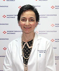
Prof. Ayşe AKTAŞ, M.D. (Head of department) (Ankara)
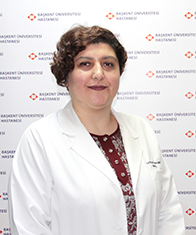
Prof. Esra Arzu GENÇOĞLU, M.D. (Ankara)

