CT Angiography
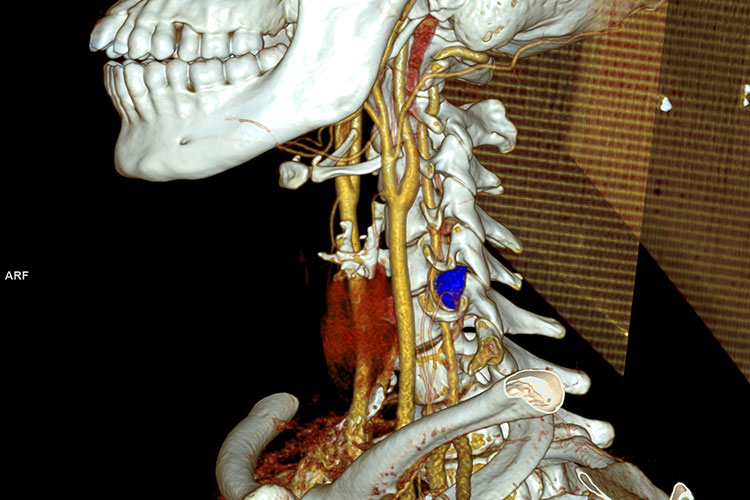
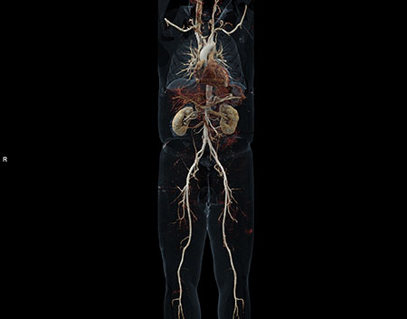
CT angiography is a technique which is used to evaluate the main vascular structures using intravenous contrast agents. Most frequent areas where CT angiography is used are coronary vessels, main neck vessels, pulmonary vessels, liver, kidneys, brain vessels, aorta and extremity vessels. CT scan is performed according to the time for the intravenous contrast agent administered via upper extremity vein to reach the target organ. Now CT angiography can be performed much more faster and higher resolution images can be obtained via multislice CT. Another advantage is that 3D images almost similar to catheter angiography can be acquired by using special computer programs.
By the Siemens Somatom Force Computed Tomography device (384 slices) which came in to service this year we can make CT and CTA scans with using less contrast material, giving lower radiation doses in shorter time when compared to the older CT devices. With the shortening of the time of scan children, un consciousness patients can be scanned without anesthesia.
Contrast agent is a substance which makes vessels become visible under x-ray. The same agent is used in cathether angiography. Common side effects of this contrast agent are flushes, metallic taste in the mouth and urinary urge. These complaints last for a few seconds. Other well known side effects are nausea, vomiting and allergic reactions. Serum creatinine levels must be checked before the examination in patients who may have kidney disease, diabetes and also old patients.
Diseases which may be diagnosed with CT angiography are aneurysms, atherosclerosis, tears in vessels called dissections, narrowing and obstructions. Follow up of “stented” patients can also be done with CT angiography.
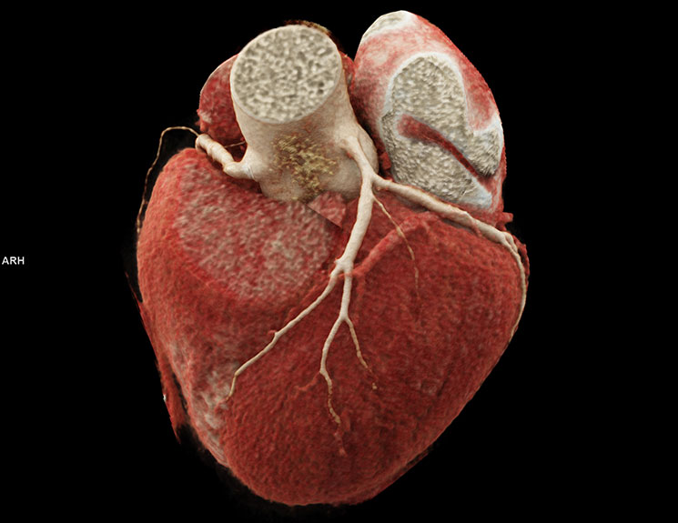
CORONARY CT ANGİOGRAPHY
This is a method which evaluates coronary arteries which supply blood to the heart by using intravenous contrast agents.
Coronary heart disease is one of the most common reasons of death. Every year in US 1,1 million of people die due to heart attack. Well known causes of heart disease are genetics, smoking, obesity, high cholesterole levels and diabetes. Another common reason is occlusion in coronary arteries because of arterial stiffness. Serious narrowing or occlusion in arteries results in heart attack because heart muscles can not be supplied with adequate blood. Sedantary life style, stress and fast food are accounted for the recent increase in heart attack rates.
In US, most of the patients admitted to the hospital with chest pain undergo catheter angiography and mostly the results are normal, which means that a lot of people are subject to unnecessary angiography. Catheter angiography is still a gold standart and it can be used for treatment as well as diagnosis but it has its own complications. Thromboembolism, dissection, aneurysm and bleeding in intervention zone are some of the complications. Thus patients with medium-high risk can also be evaluated with CT angiography. This patient group include heavy smokers, patients with family history, diabetics, and patients with hypertension. Moreover patients with chest pain who have suspicious stress test results, or suspicion of coronary artery anomaly should also be evaluated with CT angiography. This method can also be used for follow up of patients with by pass surgery.
Precise evaluation and right diagnosis is dependant on the quality of images which is in turn depends mostly on low and steady pulse rate, normal heart rhytm, adequate breath holding and patient’s cooperation during the examination. To keep the heart rate in desired range beta blokers can be used. Patient lies supine on CT table. For heart vessels to be dilated during the examination sublingual nitrogliserine is used.
Scan is performed while patient holds his/her breath after injection of intravenous contrast agent via arm vein. Scan duration is pproximately 10 to 20 seconds. Obtained images are evaluated by radiology experts.
Coronary arteries are evaluated for atherosclerosis, narrowing or obstruction caused by plaques and the amount of obstruction. The need for treatment and treatment options are considered. CT coronary angiography is also helpful for follow up of patients with by pass surgery and evaluation of stent and graft patency.
All types of CT angiography are successfully performed with multislice CT in Başkent University Ankara Hospital Radiology Department. In Ankara hospital by the new Siemens Somatom Force (384 slices) CT device all CTA procedures especillay coronary CTA are performed with lower radiation doses, shorter scan time, lower contrast material when compared to older devices. By Somatom Force coronary CTA can be performed even with high pulse rate and arrhythmia.
 |
 |
 |
Department Doctors
-
Prof. Ahmet Muhteşem Ağıldere, M.D -
Prof. Fatih Boyvat, M.D -
Prof. Kemal Murat Haberal, M.D -
Prof. Mehmet Coşkun, M.D -
Prof. Nihal Uslu, M.D -
Prof. Tülin Yıldırım, M.D -
Assoc. Prof. Özgür Özen, M.D -

Asst. Prof. Hazal Selvi Çubuk, M.D -

Asst. Prof. Burak YAĞDIRAN, M.D -
Asst. Prof. Tolga Zeydanlı, M.D -
Muhammet Kürşat Şimşek, M.D -
Nimet Akın, M.D -
Tijen CANKURTARAN, M.D