Tomosynthesis and Digital Mammography
•In the western hemisphere 1 out of 8 women is going to meet breast cancer in their lifetime. Breast cancer also can occur in men, but much more frequent in women with approximately 100 times higher.
•The most important factor for decrasing mortality ratio is the early detection. Early diagnosis is possible with regular screening and breast examinaton. Radiological examinations are very important in diagnosis.
•Mammography is the most important imaging technique for scanning and early diagnosis in the detection of breast cancer and recommended to be done regularly to women who is over 40 years old. It is not a scanning method for men, but used for early diagnosis in case of clinical suspicion. Even in the absence of a mass,by showing the calsification foci, breast cancer can be recognized in the early term with this imaging technique.
•Mammography can be applied in two ways: Analog mammography and digital mammography. In the analog mammography the image of breast is directly exposed on the radiograph. After that time, there is no possibilty for the modifying the image such as magnification and dose adjustment. Because of this reason, the suitable conditions are mandatory while imaging. In case of any need to assess any particular location in the breast repating the imaging will be required which will result in more exposure to radiation. Besides, it is not always possible to provide optimal conditons for keeping the images in long term periods and there is always risk for deterioriation or missing of the images.
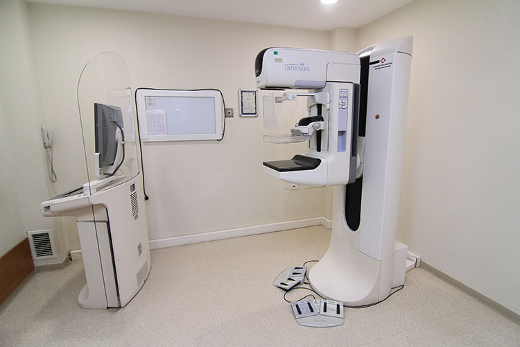
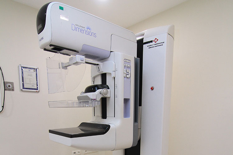
•Currently, the preferable technique for imaging of breast is digital mammography and the digital mammography images not exposed on the radiograph but transmitted to the computers. Digital mammography is a technique which uses digital (numerical) technology. After obtaining the images, in case of a need for extra investigation, the chosen area can be magnified and examined in detail and with that, there is no need for additonal images. By the changing the contrast of breast tissue, every foci in the breast can be viewed in detail.
With digital mammography, radiation dose and breast compression is decreased by calculating the thickness and density of the breast and this provides less pain which is caused by pressure on breast and also lower radiation exposition. No requirement for additional images decreases radiation level. Besides, with this method archiving is much more simple and this provides repeated usage of images in follow-ups. The risk of losing or detoriation of images are eliminated.
•But in very dense breast, even digital mammography is not sufficient for differentiating the superimposing tissue. In this situation there is a need for additional images. To eliminate this undesirable result, the last development in the breast radiology is digital mammography and tomosynthesis. Tomosynthesis is a digital mammography method which provides 3D evaluation of the breast tissue. The main difference of tomosynthesis device from the current digital mammography devices is that while the X-ray tube within the digital mammography devices is fixed, within the tomosynthesis devices the X-ray tube is mobile and this results in obtaining more than one image at a time. While with digital mammography 2D examinations are possible, the tomosynthesis provides volumetric 3D examination. Thus, the visualization of the mass which is hidden under the normal tissue becomes possible. Recent researches show that tomosynthesis can show the tumors and the borders of the tumors in the dense breast better. It’s known that the X-ray exposure in tomosynthesis is almost the same with digital mammography. The tomosynthesis requires the breast compression as with the digital mammogrphy. Currently, there is no mammography system without breast compression.
•Neither analog and digital mammography nor tomosynthesis systems can’t evaluate the internal structure of the masses. The fluid or solid chracteristics of the masses can’t be evaluated by these methods. To understand this, ultrasonography is needed.
 |
 |
Department Doctors
-

Prof. Ahmet Muhteşem Ağıldere, M.D -
Prof. Fatih Boyvat, M.D -
Prof. Kemal Murat Haberal, M.D -
Prof. Mehmet Coşkun, M.D -
Prof. Nihal Uslu, M.D -
Prof. Tülin Yıldırım, M.D -
Assoc. Prof. Özgür Özen, M.D -

Asst. Prof. Hazal Selvi Çubuk, M.D -

Asst. Prof. Burak YAĞDIRAN, M.D -
Asst. Prof. Tolga Zeydanlı, M.D -
Muhammet Kürşat Şimşek, M.D -
Nimet Akın, M.D -
Tijen CANKURTARAN, M.D