Magnetic Resonance Imaging
WHAT IS MAGNETİC RESONANCE IMAGING (MRI)?
Magnetic Resonance Imaging is a painless diagnostic method which helps your doctor in your diagnosis and lead the treatment and it usually does not require preparation. Magnetic resonance imaging uses strong magnetic fields and radio waves to create images of the organs, soft tissues, bones and other internal structures and therefore does not involve radiation used in examinations such as radiography and computed tomography. These images are then evaluated by radiologists on a computer screen and can be transmitted electronically, printed or copied to CD.
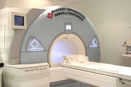
How does MRI device work?
The patient is placed inside a large magnet with a strong magnetic field and radiofrequency waves are sent on. Radiofrequency waves stimulate the hydrogen atom protons in the body. The signals that arise from the stimulated protons are collected by special receivers and body cross-sectional images are processed by a high-capacity computer. In this way, the body parts of different plans (from right to left, front to back, top to bottom) can be shown in thin sections In which cases MRI is done?
MRI is an extremely helpful method in soft tissue disease that can not be evaluated well enough with other imaging methods. In particular, in diseases of the brain and spinal cord, head and neck region and musculoskeletal system-skeletal system, MRI is the most advanced cross-sectional imaging. Also it is used safely in abdominal organs such as liver, pancreas and cardiovascular system.
What are the advantages and disadvantages of MRI?
Advantages
- There is no radiation in MRI, therefore there is no concern about the exposure to the side effects of radiation.
- Brain, heart, liver, spinal cord, soft tissue such as muscle are evaluated in more detail than other imaging methods.
- In addition to the anatomical structure, functions of the organs are examined with MRI.
- The risk of allergic side effects of MRI contrast agent is less than the risk of side effects of contrast agents used in X-ray and computed tomography.
- MRI provides a fast option in diagnosis of heart and cardiovascular system diseases without side effects.
- MRI examination is a very effective method in the diagnosis of cancer.
Disadvantages
- MRI procedures should not be done unless absolutely necessary to people with pacemakers and metal medical instruments sensitive to magnetic field and these patients should be evaluated with alternative viewing methods.
- An undetected metal object in the body affected by strong magnetic field, can cause damage to patient.
- MRI is a safe method; though there is not enough information about mother and fetus safety so in the first 12 weeks of pregnancy it is not available unless absolutely required. The second and third trimester, MRI can be done if necessary.
MRI safety
Because of high magnetic field, MRI is not eligible in some cases. If you have any of these conditions below, you are required to notify the MRI technician before imaging.
In this case, your appointment may be cancelled or MRI can be performed using special techniques.
- Pacemakers
- Neurostimulators
- Aneurysm clips
- Artificial heart valves
- Vascular graft or stent
- Drugs, such as insulin pump infusion set
- Cochlear implants (inner ear prostheses)
- Metallic implants or prostheses
- Except these situations, if you have claustrophobia, if you've worked in metal works, if there is shrapnel or bullet wounds in your body, if you have kidney disease, if you are pregnant or pregnancy is suspected and breastfeeding you should inform the technician for your own safety.
- If you had allergic reaction to contrast agents containing gadolinium in the past
MRI examinations, you should inform the technician.
Some patients may develop claustrophobia during imaging. In this case, sedative medication can be helpful. Metals, shrapnel and bullet fragments moving in a magnetic field have the possibility to harm the patients. Kidney disease is normally nota contraindication for MRI but when contrast material administration is necessary, blood tests are needed to ensure that the patient’s kidney functions are sufficient to excrete the contrast material.
MRI is not used in the first 12 weeks of pregnancy it is not used unless so required because there is not enough information about mother and fetus safety.
Paramagnetic contrast agents should not be given to pregnant women.
Unenhanced MRI has no harm in breastfeeding women. When studies with contrast conducted with lactating women, breastfeeding is asked to be stopped for next 24-48 hours after examination because the drug is passed to the baby with milk.
Tattoos and permanent make-up may disrupt the MR images. Cosmetics such as eye shadow should not be made up on the examination day because it contains metal particles. Before examination you will be asked to remove all objects such as removable dentures, hairpins, jewelry, eyeglasses, hearing aids that can disrupt MR images. Keys, coins, wallets and credit cards should be left in the locker room. You may be asked to remove your clothes and put on hospital gowns to be sure that there is no material on you that may disrupt images.
What must you do before the MRI?
In general, MRI does not require special preparation. However abdomen MRI is recommended to be performed 6-8 hours after fasting. Unless otherwise stated there is no problem to take the drugs that you use constantly. It is advised not to choose clothes containing the metal components as described above before arriving because in this case you may be requested to put on hospital gowns.
How will the MRI be done and what will you feel?
Once you get in the MRI room, MRI technician will ask you to lounge on a movable table.
After the region of the body that is desired to be examinated is positioned to the center of the cylindrical form of the device, a coil shaped receiver device can be placed on you. This coil is designed so as not to disturb you. You will hear a clicking style noise during the examination. To reduce this noise, you can wear headphones and music can be given . The technician follows you during imaging and can hear you when you say anything, so communication with the outside is provided from the the technician console. It is normal to feel the heat in the region of the body examined, but it must be reported to the technician if annoying. You are asked to stay stil in givenposition. Some patients find it uncomfortable but images must be taken when there is no movement. In some special examinations you may be asked to hold your breath.
MRI duration may vary according to the type of the requested examination. It is possible to estimate the duration before examination but this period may also vary from patient to patient. Some examinations are directly made with contrast agent but in some examinations contrast is given only when needed. In this case, images are acquired giving a gadolinium contrast agent by vascular access from your arm vein.
What will I do after the MRI?
If the contrast agent is given, drinking water can facilitate the excretion of contrast material from your body. Allergic reactions to the contrast is extremely rare. However, symptoms such as shortness of breath, skin rash and itching, should be reported immediately to technicians, and after leaving the hospital, if these symptoms occur, you should contact the nearest hospital.
You should not drive if sedatives are given because of claustrophobia. Except these situations MRI does not require special attention, you can return to your normal activities. The written report of the examination will be delivered to your doctors as soon as possible to relax you and for the quick treatment.
 |
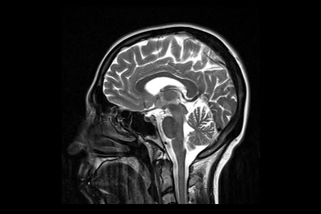 |
| Beyin MRG |
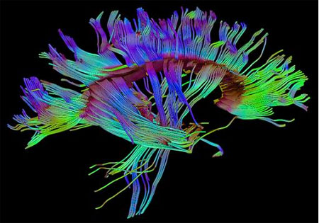 |
| Difüzyon Tensör Görüntüleme |
Department Doctors
-

Prof. Ahmet Muhteşem Ağıldere, M.D -

Prof. Fatih Boyvat, M.D -

Prof. Kemal Murat Haberal, M.D -

Prof. Mehmet Coşkun, M.D -

Prof. Nihal Uslu, M.D -

Prof. Tülin Yıldırım, M.D -

Assoc. Prof. Özgür Özen, M.D -

Asst. Prof. Hazal Selvi Çubuk, M.D -

Asst. Prof. Burak YAĞDIRAN, M.D -

Asst. Prof. Tolga Zeydanlı, M.D -

Muhammet Kürşat Şimşek, M.D -

Nimet Akın, M.D -
Tijen CANKURTARAN, M.D